


Osteochondrosis in the lumbar region is a disease that deforms and destroys the cartilaginous tissue of the intervertebral discs in the lower back.Without a layer of cartilage, the distance between the vertebrae is considerably reduced.And with the smallest sharp turns, they can change.The main danger of the disease is the possibility of forming an intervertebral hernia.
Can't you look to raise an object that has fallen to the ground?Do you suffer from acute pain in the lumbar column and go often, wrapping the size in a hot scarf?Do not ignore the condition that bothers you.
Osteochondrosis in the lumbar region can drag in its duration for a long time.There is no need to discover the body for strength.Love your body.And he will reverse.
The lumbar region represents most of the burden of all body weight compared to the departments of the chest and the cervix.Consequently, this subspecies of osteochondrosis is the most common.
What are the stages of the development of osteochondrosis?
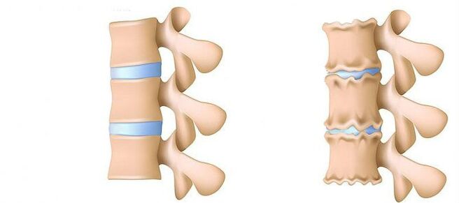
- 1st step.Precision.The height of the disc is reduced.In the fibrous ring (the outer layer of the intervertebral disc of the cartilaginous fibers), a crack is formed.Lumbar muscles are starting to get tired quickly.You feel a certain discomfort in the back.
- Step 2. Violations of metabolic processes in the nucleus jacket (central part of the intervertebral disc, which consists of a cartilage jacket): its cells are dead or completely destroyed.The structure of collagen (the structure of proteins is based on connective tissue) of the fibrous ring is also disturbed.Local pain, a person cannot face the physical activity he previously considered quite doable.
- STEP 3. Complete destruction of the fibrous ring.The adjacent vertebrae cease to be stable.Any uncomfortable installation causes pain.Due to the experience of the nerve roots which move away from the spinal cord, the members can become less sensitive and mobile.
- 4th step.The tissues of the intervertebral disc become scar.The vertebra can be in the shell.The clinical description here depends on individual physiology.
Lumbar pain (lumbago) and the pain that gives the leg during the sciatic nerve (ISHIAS) is one of the most common complaints that patients are looking for a doctor.Due to the fact that these symptoms are quite common in the general population and that their regular growth is also noted, the diagnosis and treatment of these patients will remain one of the main areas of activity of neurosurgical hospitals.Despite the widespread of this pathology, the surgical elimination of the hernia of the intervertebral disc (MPD) is only required in 10% of patients with the clinical image of the lumbar algia.In the remaining part of the patients, the best effect has conservative treatment, including the drug, physiotherapy exercises, the use of physiotherapeutic treatment methods, as well as a return to previous daily physical activity.
Steps of the disease
Degenerative-dystrophic processes most often start with deterioration of the function absorbing the shocks of the intervertebral disc.
- Damage of blood supply to the intervertebral disc.In adults, the food of the intervertebral discs is carried out by the diffusion: the blood is only delivered to the vertebrae, and already through them, it "infiltrates" in the discs.In the best way, the disc is powered during dynamic loads (for example, walking), because the principle of the pump (outlet of the transformed fluid when compressed, the flow of nutrients and oxygen when removing the load).Thus, the nutrition of intervertebral discs is difficult in particular under the conditions of a sedentary lifestyle (hypodynamia).
- Changes in the pulp disc kernel.With a deterioration in blood supply, water supply, sugars and amino acids with a pulpose nucleus is disrupted.For this reason, the production of carbohydrates connecting water.The nucleus is dehydrated, its gel -shaped structure turns into a fibrous, the ability to spring and extinguish the shots gets worse.This increases the load on the fibrous ring and the vertebrae, they are more likely to be blocked and injured.
- Changes in the fibrous ring of the intervertebral disc.Due to the flattening of the pulpose nucleus, the increased load is on the fibrous disc ring.In conditions of poor blood diet, the fibrous ring loses its strength.The instability of the spine occurs, which can lead to the formation of an intervertebral hernia, a displacement of the vertebrae and damage to the spinal cord or the nerve roots.
- Disk protection.The formation of an intervertebral hernia.While the fibers of the fibrous ring weaken, the Pulpian nucleus begins to come out, for example, to the intervertebral channel (protrusion of the disc).Such an amazing can still lead to a break in a fibrous ring and the formation of a hernia.Find out more about the training process of an intervertebral hernia in a separate article - "effective treatment of intervertebral hernia at home".
- Spondylosis is the destruction of the intervertebral joints (spondylartrosis), the growth of osteofites and the ossification of ligaments.In addition to the formation of an intervertebral hernia in osteochondrosis, damage to intervertebral joints, destructive changes in the vertebra (cartilage) and ligaments are observed.
As osteochondrosis and the development of complications are progressing, you should use more and more often increasing doses.This leads to high financial costs, as well as a new deterioration in health due to the side effects of drugs.
Pharmacotherapy, as a rule, is supplemented by the immobilization of a friend of the spine by using orthopedic corsets of variable degrees of stiffness.
Surgical treatment is justified only in cases where the level of compression of the vertebral rook, determined by the clinic, corresponds to the examination confirming the rupture of the fibrous ring with the "loss" of the hernia of the MPD in the light of the vertebral canal [3–6].The results of the surgical treatment in patients with small disk protuberances, as a rule, are disappointed with the doctors and the patient himself.The method for establishing a precise diagnosis is magnetic resonance imaging (MRI).About 10% of people in a common population are impossible to lead a routine MRI due to claustrophobia (fear of closed spaces).In this category of people, it is possible to use the "open" MRI if called, with the corresponding image quality loss obtained.Patients who have already undergone surgical treatment are necessary to carry out an MRI with a contrasting strengthening to delimit the postoperative changes of the scar from the real hernial projection of the disc.In patients suspected of MPD hernny projection, when the implementation of MRI is impossible, or the results obtained are non -informative and calculated tomographic myelography (CT) acquires a particular diagnostic value.
Private diagnostic specialists who interpret the results of studies, as a rule, exaggerate the degree of damage to disc due to the impossibility of comparing clinical data with "discoveries" during tomography.Conclusions such as "the changes correspond to the age of the patient" are almost never found in research protocols.Despite the improvement of neuroimaging techniques, the responsibility for the properly deceived diagnosis is the shoulders of the clinician, as he can only compare the clinical image with the data obtained during tomography.An increase in the resolution of tomographies has slightly improved the results of surgical treatment, but the differences in relation to the standard in asymptomatic patients have started to be detected.The process of process accompanying the degenerative-degenerative lesion of the spine has undergone serious progress in the latestyears.Arthropathy of the arched joints is widespread in the general population and is detected quite often in people in the middle and older age group during research CT. Degenerative changes in MPD, which are also widely used, are often detected and MRI is a more specific method for their diagnosis.At the same time, the changes pronounced in the MPD are not uncommon, not accompanied by a rupture of the fibrous ring, but were manifested only by a slight "stab" of the disc in the light of the vertebral canal or the intervertebral holes.In some cases, the degenerative processes occurring in the MPD can cause the destruction of the fibrous ring with subsequent ruptures, which causes the migration of part of the pulp nucleus outside the disc with the compression of the adjacent roots of the spinal cord.The assertion that if the pain in the leg is noted, it must necessarily be raped on the roots of the spinal cord is not entirely true.Pain in the buttock with irradiation on the posterior surface of the thigh, can lead to both the degeneration of the MPD itself and the arched intervertebral joints.For a real Ishialgia attack caused by the compression of the koreshka of the nerve of the MPD hernia, the pain radiates on the posterior surfacethigh and lower leg.Undeterminated pain, limited only to the glued area or the thigh area without distribution along the sciatic nerve, as well as bilateral pain in glued areas or hips that change their location (either right, then left), are more often caused by arthropathy of arched joints or diffuse degeneration of MPD.The clinical image of Koruska compression of MPD hernia can also be a concomitant pathology (for example, osteoarthritis of the knee joints).In patients with such pain, surgical treatment will not have the appropriate effect, regardless of the pathology detected by tomographic examination.In other words, in patients only with the pain clinic in the back, the elimination of MPD hernia will be ineffective, even if the tomograms are determined by the projection of the MPD, as usual and occurs.But there are also patients in whom the typical Ishia image is accompanied by pronounced invalid pain syndrome, while during studies carried out using very perceptual tomographies, the compression of the roots of the spinal cord is not determined.This category of patients is inappropriate to carry out surgery, because over time, the radicular symptoms, as a rule, are calm.
It is necessary to clearly imagine the mechanisms leading to the development of the hernial projection of the MPD in order to recommend to patients the volume of movement authorized, without forgetting the work activity.The forces which contribute to the formation of the hernial projection are the result of degenerative changes in the MPD and a decrease in the vertical (height) of the fibrous ring and the pulpoose nucleus.The slot fragment of the MPD in 80% moves in the posterior direction, while introducing in the light of the vertebral canal and the medial sections of the intervertebral hole.This displacement of the hernia from the MPD to the midline is facilitated by the strength of maintaining the posterior longitudinal ligament.Up to 10% of hernial provisions are localized laterally and spread to the intervertebral hole (Forsin Hernias) or to the outer edge of the hole where the cerebrospinal spine leaves it, tightening it.
In the process of vital activity, dehydration and degenerative changes cause loss of the height of the MPD.These pathological processes involve both a fibrous ring and a luscious nucleus.The most pronounced destruction of the pulpose nucleus in the backdrop of concomitant degeneration of the fibrous ring, as a rule, only leads to the loss of the height of the MPD without its significant gatherings.With predominant changes in the fibrous cycle, the vertical forces affecting the pulp nucleus preserved and which are a derivative of their own weight, as well as the back muscles, acting on the disc in the lateral direction, exerts excessive pressure on the remaining fragment of the pulpoose nucleus, which cannot keep the fibrous ring in place.
The summons of these two forces leads to an increase in centrifugal pressure on the MPD, which, with the stretch component acting on the fiber of the fiber ring, can cause its rupture and its fragment of fragments of the nucleus of the remaining pulp.After the formation of an hernial projection and that the "redundant" fragment of the Pulpian nucleus was outside the fibrous ring, the structure of the MPD becomes stable [2].Following the forces affecting the nucleus and the altered degeneratively altered cycle of the MPD, they are balanced and their vector, which contributes to the pursuit of the projections of the fragments of the nucleus, fades.In some cases, the partial degenerative changes of the pulpos nucleus contribute to the formation of gas inside the MPD, followed by excessive pressure on its remaining fragment.The formation of a hernia is also accompanied by the gas formation process inside the disc.
An excessive and strong physical activity shown on the patient's back, in the context of the existing-dystrophic degenerative lesion of the vertebral column, is generally only a trigger which leads to a detailed clinical image of a compression radicular syndrome, which is often and erroneous by the patients themselves, like the primordial of the lumbar.Clinically, MPD hernia can manifest itself with reflex and compression syndromes.Syndromes are referred to compression, in which the hernial projection is drawn, pressed and deformed, the blood vessels or the spinal cord are compressed and deformed.Reflexes reflexes include syndromes caused by the effects of disc hernia on the receptors of these structures, mainly the end of the vertebral nerves, which leads to the development of reflex and tonic disorders manifested by vasomotor, dystrophic and myofascial disorders.
As indicated above, surgical treatment with a degenerative-systrophic lesion of Posvinor is only advised in 10% of patients, the remaining 90% react to conservative measures.The basic principles of the use of these are:
- pain syndrome relief;
- restoration of the correct posture to maintain the fixing capacity of the modified MPD;
- elimination of muscle and tonic disorders;
- restoration of blood circulation in the roots and the spinal cord;
- normalization of conductivity in nerve fibers;
- elimination of scar and spacing changes;
- Relocation of psycho-somatic disorders.
Treatment
Today, in the treatment of osteochondrosis and its complications, drugs from the following groups are used:
- Net -Ear anti -inflammatory (NSAIDs) -in the form of tablets or drug injections.These funds have the capacity to reduce pain, reduce the activity of inflammation.However, the effect of their use does not last long - from several hours to two to three days.Therefore, these funds must be taken for a long time - weeks, and sometimes months.At the same time, these drugs negatively affect the mucous membranes of the gastrointestinal tract.Their long -term reception is heavy with the development of gastritis, ulcerative lesions.In addition, they can negatively affect the work of the kidneys, the liver and contribute to the development of hypertension.And, at the same time, these funds do not contribute to cleaning the discs of dead cells.Therefore, their use is just a way to relieve symptoms for a while, but not to eliminate the main problem.
- CTEPOID anti-inflammatory drugs (Gopmonal).As a rule, they are used for severe and impenetrable pain accompanying hernias, radiculites, ishias, etc.Gopmons have the capacity to eliminate manifestations from inflammation (due to the oppression of the immune system), relieves pain.But they also negatively affect the mucous membranes of the stomach and the intestines, promote the leachate of the calcium of the bones, inhibit the production of their own vican gopmons.And do not contribute to cleaning the attention of dead cells.
- Papasmolics are drugs that affect muscles or nerves that go to muscles and cause the relaxation of skeletal muscles.These means help relieve muscle pliers for a while, reduce pain and improve blood flow.But at the same time, they do not help clean the tissues of dead cells.Therefore, they do not help heal osteochondrosis.
- EPIDUPPAL BORDAUDE - The introduction of pain relievers and gopmonal agents in space between the solid brain shell and the perioste covering the vertebrae.It is used, as a rule, for intense pain - in the acute period of intervertebral hernia, with severe radicult, Ishias.Depending on the composition, such an injection helps relieve pain for several hours to several days.After the expiration date, the manifestations of the disease are returned, because the procedure does not help to restore metabolic processes in the discs.In addition, when carried out, there is a risk of injury to blood vessels and nerves.
Conservative treatment methods include various orthopedic effects on the spine (immobilization of the corset, traction, manual therapy), physiotherapy (therapeutic massage, physiotherapy exercises, acupuncture, electrotherapy, mud, different types of heating), paravertebral, epidural blockage and medication therapy.The treatment of the degenerative-dystrophic lesion of the spine must be complex and gradually.As a rule, the general principle of conservative measures is the appointment of pain relievers, non-steroidal anti-inflammatory drugs (NSAIDs), muscle relaxants and physiotherapy.
The analgesic effect is obtained by the appointment of diclofenac, ketoprofen, lornoxicam, tramadol.A pronounced analgesic and anti-inflammatory effect has Loroxes, existing both in the form of injection and tablets.
NSAIDs are the most widely used drugs for degenerative-systrophic damage to the spine.They have an anti -inflammatory, analgesic and antipyretic effect associated with the suppression of the cycloxygenase enzyme (COC -1 and TSOS -2), which regulates the transformation of arachidonic acid into prostaglandins, prostacillas, thromboxans.In the elderly and patients with risk factors for side effects, it is advisable to carry out the "coverage" of gastrotrotter under "coverage".In these patients, at the end of the course of therapy by injection of NSAIDs, the transition to the forms of tablets of the inhibitors of the COO -2, who have a lower severity of the side effects of the gastrointestinal tract, is advised.
To eliminate pain associated with the increase in muscle tone, it is advisable to include central musclex in complex therapy.
Surgical treatment of the degenerative-dystrophic lesion of the spine is justified with the ineffectiveness of complex conservative measures (within 2 to 3 weeks) in patients with MPD hernia (generally more than 10 mm) and non-loading radicular symptoms.There are emergency indications for surgery with a "abandoned" sequesttra in the light of the vertebral canal and an expressed compression of the roots of the spinal cord.The development of Caudal syndrome is facilitated by acute radiculomilohemilo, leading to severe hyperalgic syndrome, when even the prescription of drug pain relievers, the use of blocking (with glucocorticoids and anesthetic) does not reduce the seriousness of pain.It is important to note that the absolute size of the hernia of the disc has no determining value to make the final decision on surgical intervention and must be considered in relation to the clinical image and the discoveries detected by Tomographic examination.In 95% of cases, access open to the vertebral canal is used in hernia.Various discopation techniques (coagulation of cold plasma, laser reconstruction, etc.) have not currently been today, and their use is justified only for the protuberances of the MPD.The classic open -open microshrurgical elimination of the hernia of the disc is made using microsurgical tools, binocular aggressions or an operational microscope.Analysis of remote processing results (in more than 2 years) 13,359 patients who have been eliminated by MPD hernia, 6135, the sequester of which has been removed and 7,224 aggressive discs were made more likely (7% against 3.5%) in patients who only eliminate sequestration.The quality of life is more reduced in patients with pain syndrome, while the repeated formation of hernias does not always manifest itself clinically.
In conclusion, I would once again highlight the need for a thorough clinical examination and an analysis of tomograms to make an optimal decision on the choice of tactics for the treatment of a particular patient.